- Home
- under high
- Correlative light and electron microscopic observation of calcium phosphate particles in a mouse kidney formed under a high-phosphate diet
Correlative light and electron microscopic observation of calcium phosphate particles in a mouse kidney formed under a high-phosphate diet
4.5 (574) · $ 15.50 · In stock

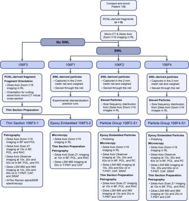
GeoBioMed perspectives on kidney stone recurrence from the reactive surface area of SWL-derived particles
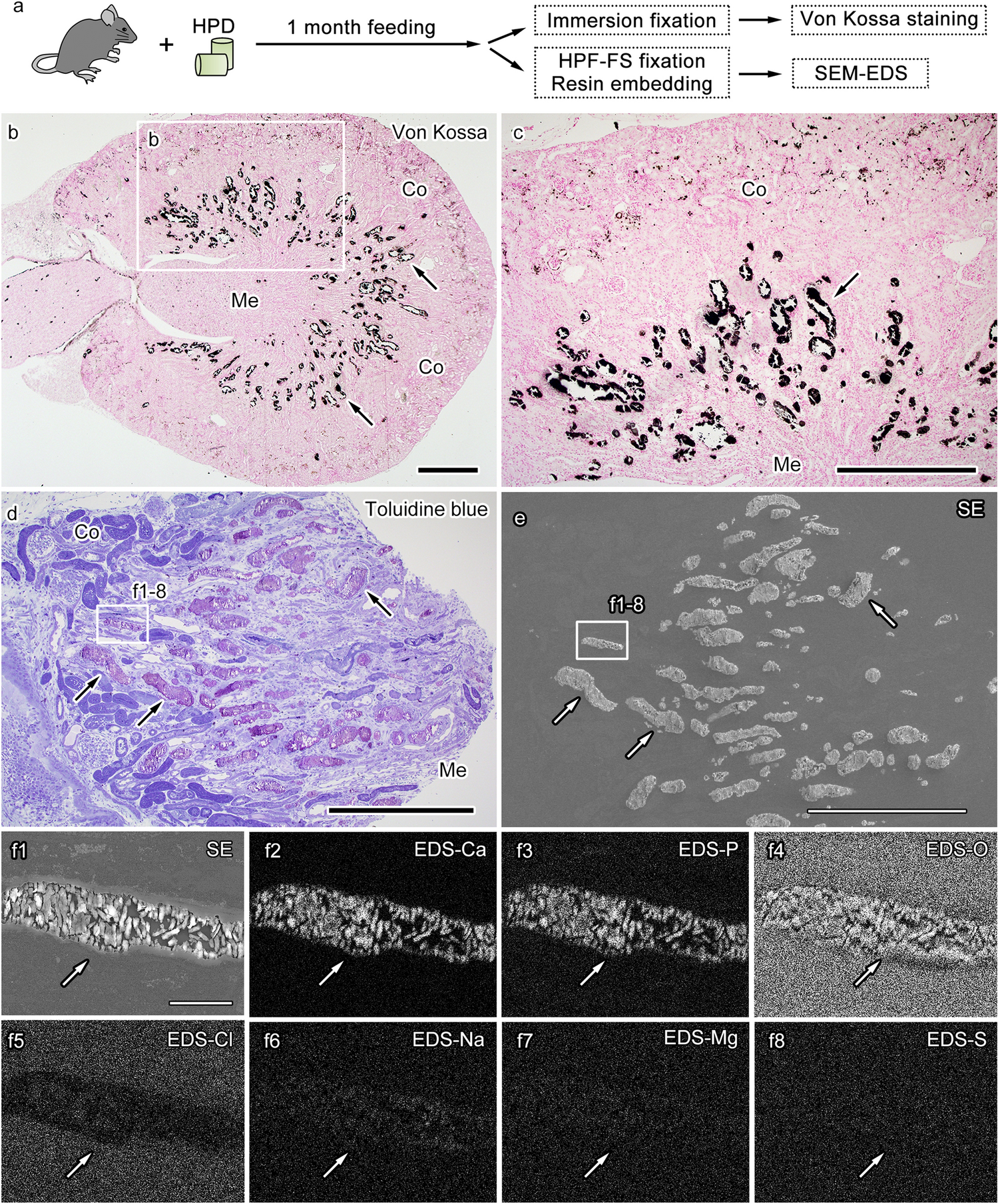
Correlative light and electron microscopic observation of calcium phosphate particles in a mouse kidney formed under a high-phosphate diet

DIVISION OF HISTOLOGY, JICHI MEDICAL UNIVERSITY, SCHOOL OF MEDICINE

DIVISION OF HISTOLOGY, JICHI MEDICAL UNIVERSITY, SCHOOL OF MEDICINE

Ultrastructure and Nanomorphology of the American Mink (Mustela vison) Kidney

Arterial Calcification in Chronic Kidney Disease: Key Roles for Calcium and Phosphate

Ultrastructure and Nanomorphology of the American Mink (Mustela vison) Kidney

b-e Figs. 7f, g Sup Fig. 6f, 11, 12 Figs. 1-3, 5a, g, m, s, Sup Fig. 1

Transmission electron microscopic and X-ray absorption fine structure spectroscopic investigation of U repartition and speciation after accumulation in renal cells

Effects of prolonged dietary phosphate load on renal IL‐36α expression.

Antibacterial and osteoinductive properties of wollastonite scaffolds impregnated with propolis produced by additive manufacturing - ScienceDirect

PDF) Characterisation of Calcium Phosphate Crystals on Calcified Human Aortic Vascular Smooth Muscle Cells and Potential Role of Magnesium