Ultrasound imaging - Download as a PDF or view online for free
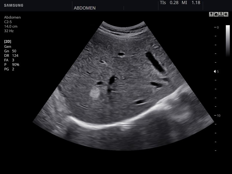
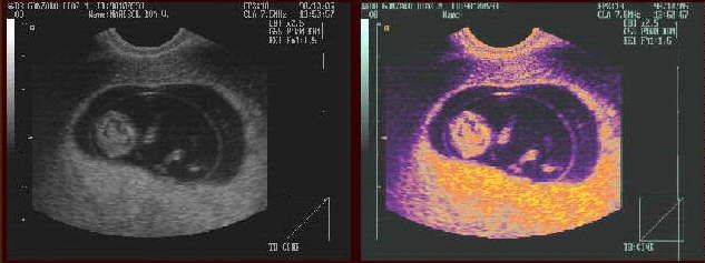
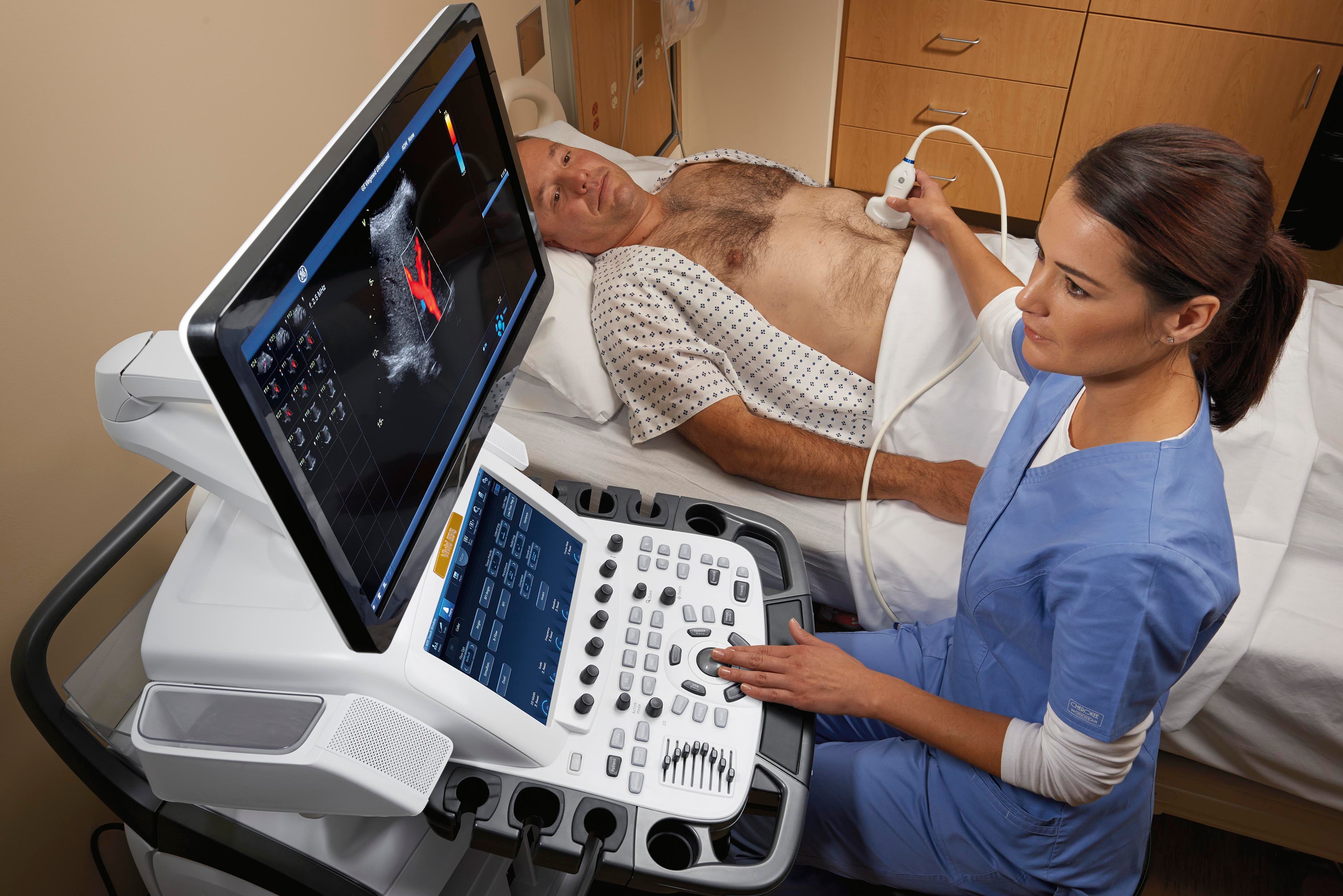
Ultrasound uses high frequency sound waves to visualize internal structures. It works by transmitting sound waves into the body using a transducer probe, which detects the echoes as they bounce off tissues and organs. The echoes are processed to form images on the ultrasound machine screen in real-time. Common applications include obstetrics, cardiology, and urology. The Philips HD11 is an ultrasound system with curvilinear, linear, and phased array probes for different exams. It provides grey scale, Doppler, and color imaging modes. Ultrasound has benefits of being non-invasive, portable, and having no radiation, but has limitations of being operator dependent and unable to penetrate bone.
Highlight, take notes, and search in the book

Radcases Ultrasound Imaging (Radcases Plus Q&A)

Ultrasound imaging results of the fiber-optic array ultrasound sensor.
Rehabilitative Ultrasound Imaging (RUSI)

Enhancing Safety with Ultrasound Imaging — Dr. Heather Friedman ND, LAc
Ultrasound 101 - Part 4: Depth and focus

Reading Minds with Ultrasound: A Less-Invasive Technique to Decode the Brain's Intentions

The Principles of Ultrasound Imaging

Through-needle all-optical ultrasound imaging in vivo: a preclinical swine study
Advancements in Ultrasound

Texas Ultrasound Imaging, LLC
What Is an Ultrasound Machine and How Does It Work? - Ultrasound Solutions Corp.

Functional ultrasound imaging of the brain