a Mandibular fistula indicated by an arrow in the apical region of dd
5 (530) · $ 11.00 · In stock
Download scientific diagram | a Mandibular fistula indicated by an arrow in the apical region of dd 36-37. b A fistula in the apical region of dd 46-47 (white arrows) and a red area in the mucosa (black arrows) are seen in the right lingual surface of the mandible. c Panoramic radiograph showing no bone lesions in the mandible. d Periapical x-ray with no bone involvement in the apical region of dd 46-47 from publication: Treatment of bisphosphonate-induced osteonecrosis of the jaws with Nd:YAG laser biostimulation | Osteonecrosis, Jaw and Nd:YAG Laser | ResearchGate, the professional network for scientists.
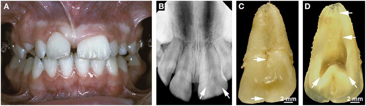
Frontiers Malformations of the tooth root in humans
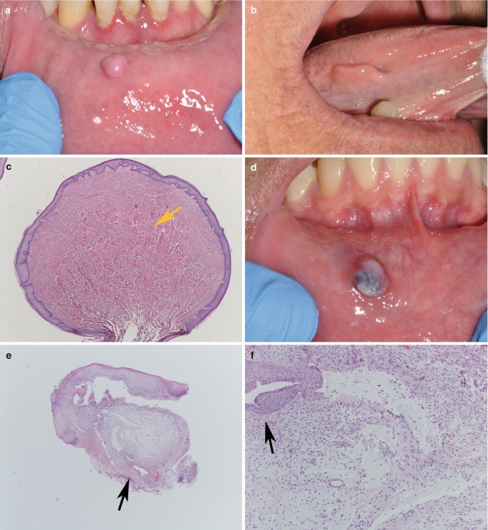
Benign Lumps and Bumps
Case Archive, School of Dental Medicine

VRF as an Endodontic Periodontal Lesion

/profile/Maria-De-Souza-3/publ

Healthcare, Free Full-Text

Ultrasonographic Imaging in Periodontology

JaypeeDigital

JaypeeDigital

a Mandibular fistula indicated by an arrow in the apical region of dd

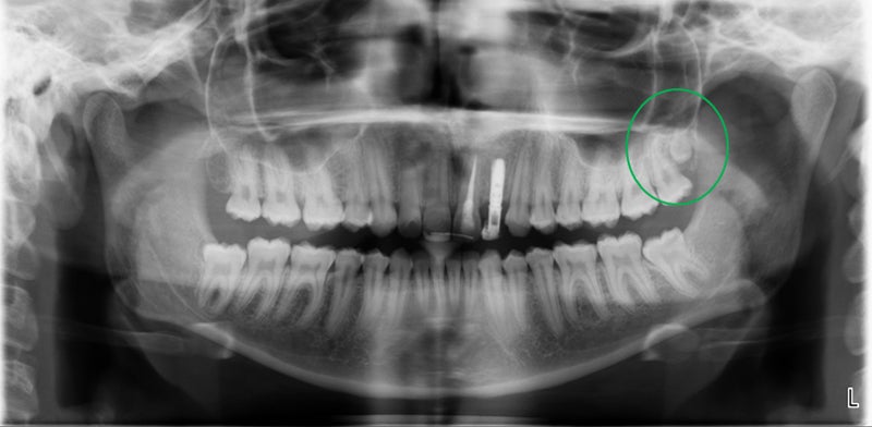
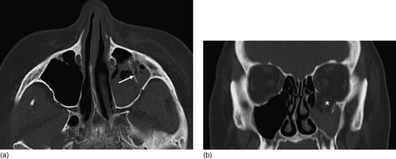
Medication-related osteonecrosis of the jaw without osteolysis on computed tomography: a retrospective and observational study
Imaging in trauma (Section 4) - Trauma