Pitfalls of inferior vena cava M-mode – NephroPOCUS
5 (529) · $ 27.00 · In stock

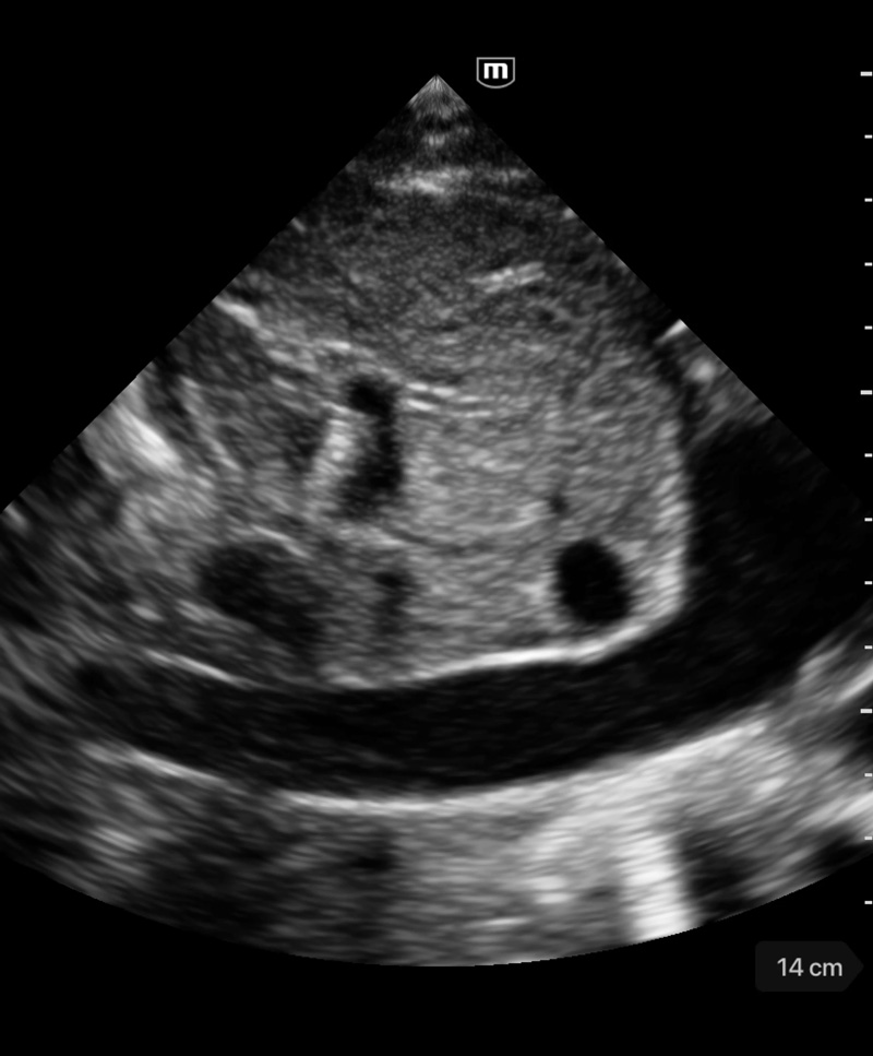
Visual estimation of IVC collapse on B-mode (grey scale image) is generally preferred to M-mode, though in theory, M-mode measurement might be able to give accurate collapsibility index. There are several reasons for this. A major limitation of IVC M-mode is that the vessel moves mediolaterally and craniocaudally during respiration, with collapse occurring off axis…

The 'ring of fire' Foley balloon – NephroPOCUS

PDF) Transcending boundaries: Unleashing the potential of multi-organ point-of-care ultrasound in acute kidney injury
JCM, Free Full-Text

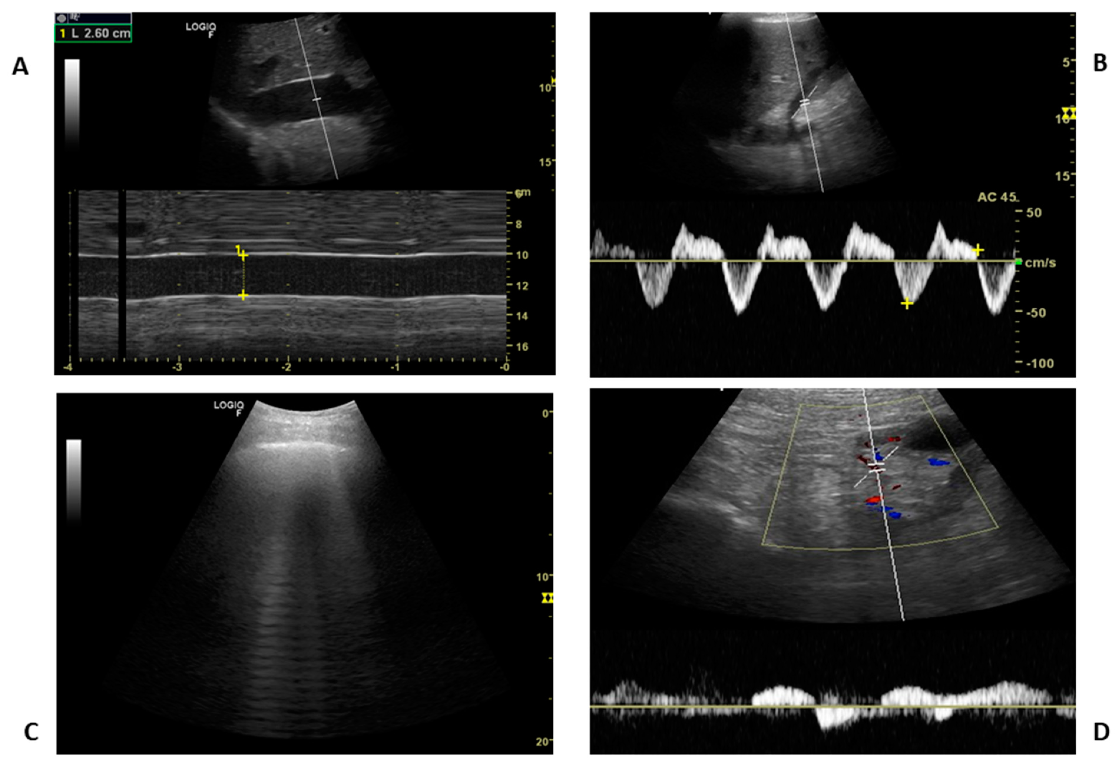
Image Acquisition Method for the Sonographic Assessment of the

The 'ring of fire' Foley balloon – NephroPOCUS

Venous Excess Doppler Ultrasound for the Nephrologist: Pearls and Pitfalls - ScienceDirect

PDF) Transcending boundaries: Unleashing the potential of multi-organ point-of-care ultrasound in acute kidney injury
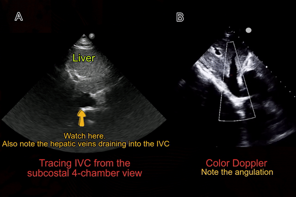
Inferior Vena Cava POCUS: The Basics of Image Acquisition - Renal

Pitfalls of inferior vena cava M-mode – NephroPOCUS

Image Acquisition Method for the Sonographic Assessment of the

PoCUS in nephrology: a new tool to improve our diagnostic skills
Inferior Vena Caval Thrombosis: Practice Essentials, Anatomy

Figure 13 from Congenital absence of inferior vena cava

Point-of-care ultrasound in diagnosis and management of congestive